Image Gallery
Gallery
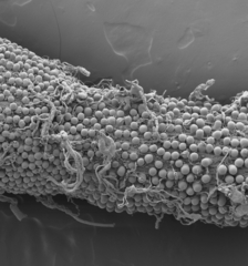
SEM of Beetle Gut
Media Details
Created 1/6/2009
This scanning electron micrograph shows the exterior surface of a mealworm beetle's (Coleoptera: Tenebrionidae: Tenebrio molitor) midgut. The surface of the midgut, which is ordinarily bathed in the insect's blood (hemolymph), attracted our attention because of the numerous pouches that protrude from it. These pouches appear on the outer surfaces of other beetle midguts, but only after the transformation from pupa to adult. Prior to metamorphosis, the outer surfaces of both the larval and pupal midgut are smooth. We are currently trying to determine why only beetles -- of all insects -- show this distinctive midgut architecture and what function these numerous pouches have at only the adult stage of the beetle life cycle.
Credits
- Mark Bee , ITG, Beckman Institute
- James Nardi , Department of Entomology