Image Gallery
Gallery
Computer Modeling of the Levator Veli Palatini Muscle
Media Details
Created 12/5/2006
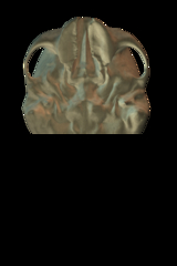
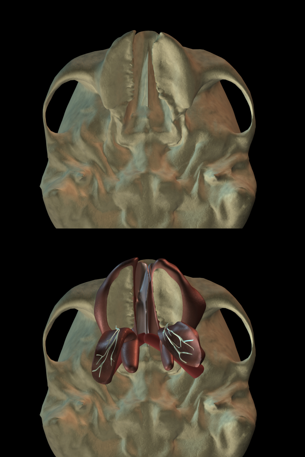
A child born with a cleft palate may undergo multiple surgeries to restore anatomy and regain muscle function. In numerous cases, even after surgical intervention to repair the cleft palate, normal sounding speech may not be restored due to improper surgical alignment of a palatal muscle known as the levator veli palatini (levator). Success in surgery is contingent upon a better understanding of the structures and functions of the levator muscle and surrounding soft tissue. The goal of the project is to demonstrate a proof of concept in the use of MRI and 3D computer technology for the purposes of presurgical planning, assessment of levator muscle morphology before and after surgery, and postsurgical assessments of the levator muscle. In this instance, the image represents a view of the undersurface of the skull with the mandible removed, and demonstrates the cleft of the palate and the surgical procedure used to reconstruct the internal anatomy. This research is a step closer to providing significant advances in the clinical realm such as reducing human error during surgery, computer simulation of surgical planning, as well as patient specific modeling, diagnosing, and treatment for individuals born with a cleft palate. This project demonstrates the implementation of translational research by combining basic research with applied clinical research.
Credits
- David Kuehn, PhD , Department of Speech and Hearing Sciences
- Michael Goldwasser, DDS, MD , Carle Foundation Hospital
- Jamie Perry, MA, CCC-SLP , Department of Speech and Hearing Sciences