Image Gallery
Gallery
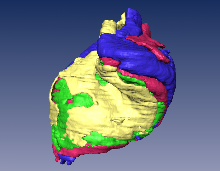
Human LGN Model

Media Details
Created 1/25/2005
The model displayed here was derived from a real human Lateral Geniculate Nucleus (LGN), the portion of the brain responsible for receiving image information from the retina. The colored regions represent four layers of the LGN, where each layer processes visual information in a slightly different manner. The different layers represent differing color components of an object in a spatial field. The layers were isolated from individual histological sections scanned on the Nikon LS-8000 (the Super Coolscan) scanner in the VMIL. The individual layers were segmented and reconstructed within the analysis package Amira, which produced the rendering displayed here.
Credits
- Janet Sinn-Hanlon , ITG, Beckman Institute
- Joseph Malpeli , Department of Psychology
- Kelly Kraft , Department of Psychology