Image Gallery
Gallery
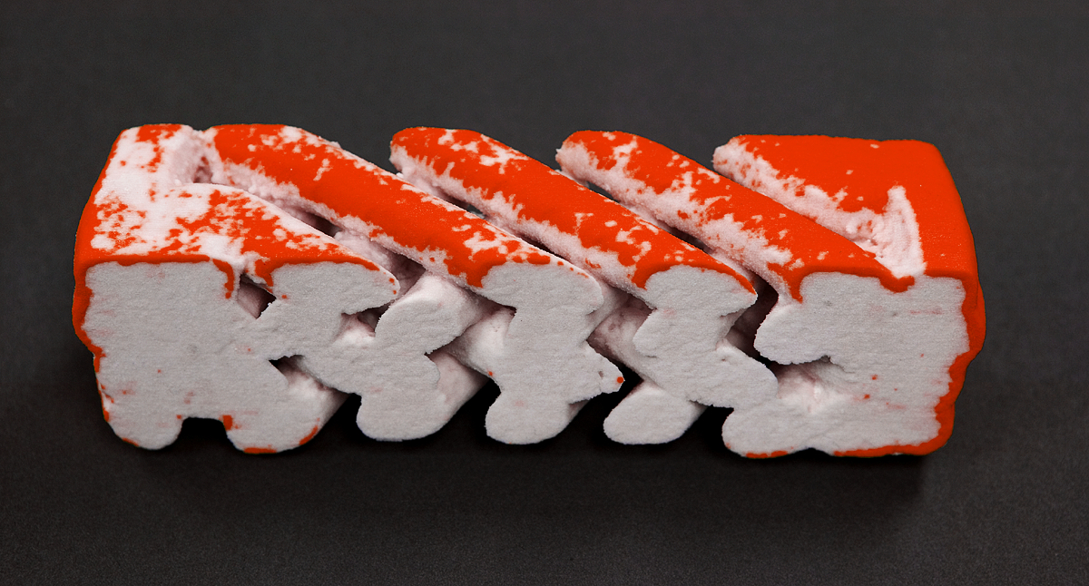
Os-Stained Tissue on a HA Scaffold

Media Details
Created 10/26/2004
Mouse D1ORL stem cells are cultured on the scaffold for 2 weeks prior to fixation (Os-staining) and micro-CT imaging. Using a raw data histogram, the tissue and the HA can be distinguished based on their x-ray attenuation values. Values associated with the tissue are assigned the color red. The width of the scanned region of the scaffold is 1.2 mm, and the model is printed at approximately 30X. Processing and visualization were accomplished in the VMIL. The 3D model was printed in plaster on the ZCorp Z406 rapid prototyper and infiltrated with cyanoacrylate. The data processing was done using Analyze. The model was photographed using the Canon 1Ds 11MP digital-SLR.
Credits
- Amy Wagoner Johnson , Mechanical Science & Engineering