Image of the Week Gallery
Leslie's Lung
Media Details
Created 4/13/2004
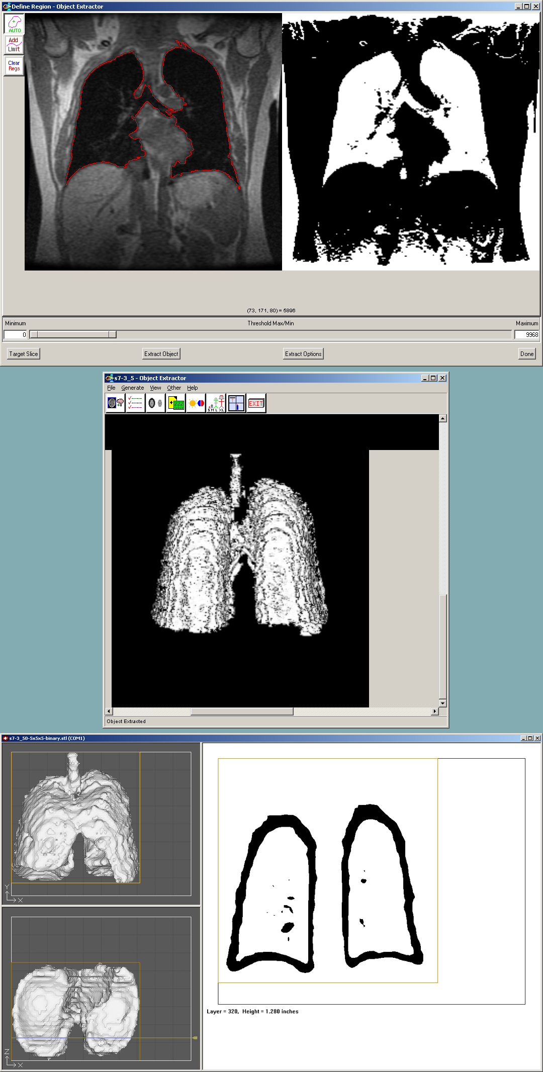
The sequence of images here demonstrate the process of converting MRI data into a solid structure to be printed on the Z406 3D printer. The first image demonstrates one step in the segmentation of the lung tissue using the Object Extractor tool in Analyze. The second image displays one view of the extracted three dimensional surface. The third image displays the model as loaded into the ZPrint software. A feature of interest in this image is the hollow nature of the lung cavity. The most recent version Analyze provides the capability of generating surfaces as "thin shells". The thickness of the shells can be specified in advance. By making a normally solid part into a thin shell, the operator of the printer can drill a small hole in the object to release any unbound powder, thereby saving money in the cost of the print. The lung will be included in the MFA Thesis Exhibition at the Krannert Art Museum April 24th 6-8pm (opening). The show will continue through May 16th. This is a series of works done by Sculpture Graduate Leslie Speicher. She has been studying the internal landscape of her body and representing its parts in a variety of ways.
Credits
- Leslie Speicher , School of Art & Design
- Daniel E. Weber , ITG, Beckman Institute