Image of the Week Gallery
Montastraea faveolata Skeleton Biopsy

Media Details
Created 2/17/2009
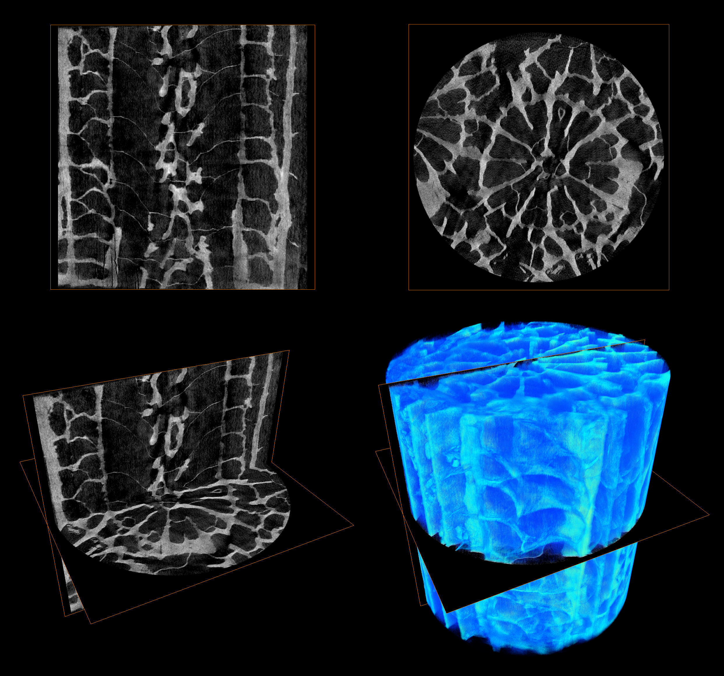
MicroCT images of a skeleton biopsy extracted from Montastraea faveolata, a reef-building coral from the fringe reef of Curacao, Netherlands Antilles. The top two images are single orthogonal slices of intersecting image planes from the 3-D microCT scan. These 2-D slices show transverse and longitudinal transects of a single coral polyp (~2mm in diameter). The bottom left image shows these slices as they exist in 3-D space. Finally, the bottom right image reveals a 3-D volume rendering using color-mapping to represent regions of varying densities in the skeleton. The images were acquired on the ITG's new Xradia BioCT in the Microscopy Suite and later visualized and reconstructed with Amira software in the Visualization Laboratory.
Credits
- Carly A. Hill , Department of Geology
- Bruce W. Fouke , Department of Geology, Institute of Genomic Biology, and Department of Microbiology