Image of the Week Gallery
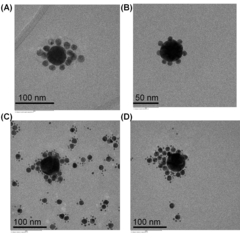
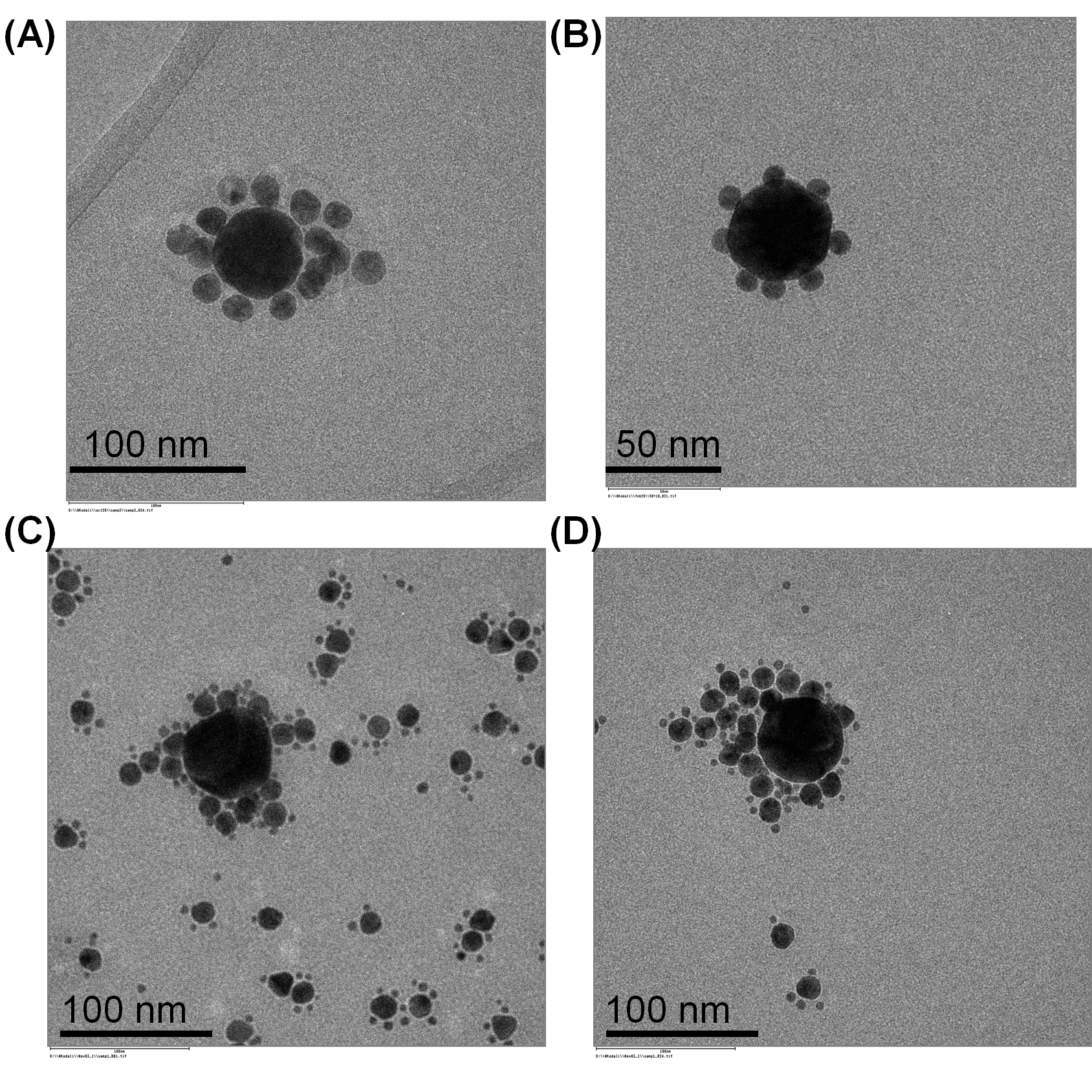
Structured Aggregates of Variously Sized Gold Nanoparticles
Media Details
Created 5/4/2010
Transmission electron micrographs of structured aggregates of gold nanoparticles of different sizes. Shown are symmetric spherical aggregates with gold nanoparticles of (A) 60 and 15 nm; (B) 50 and 10 nm; and (C,D) 60, 15, and 5 nm. The gold nanoparticles were aggregated through the linkage of synthetic oligonucleotides that were functionalized onto their surfaces. Images were collected using the Philips/FEI CM200 transmission electron microscope (TEM) in the Beckman Microscopy Suite.
Credits
- Anil K. Kodali , Department of Mechanical Science & Engineering
- Pratik Randeria , Department of Bioengineering
- Matthew Schulmerich , Department of Bioengineering
- Rohit Bhargava , Department of Bioengineering
- Laura Jane Elgass , Department of Bioengineering
- Rohun Palekar , Department of Bioengineering