Fees
Microscopy Suite Equipment Fees
Fees/rates for use of equipment in the Microscopy Suite determined through analysis by Government Costing, University of Illinois. In effect starting August 1, 2021; subject to change.
Raman Confocal Imaging System

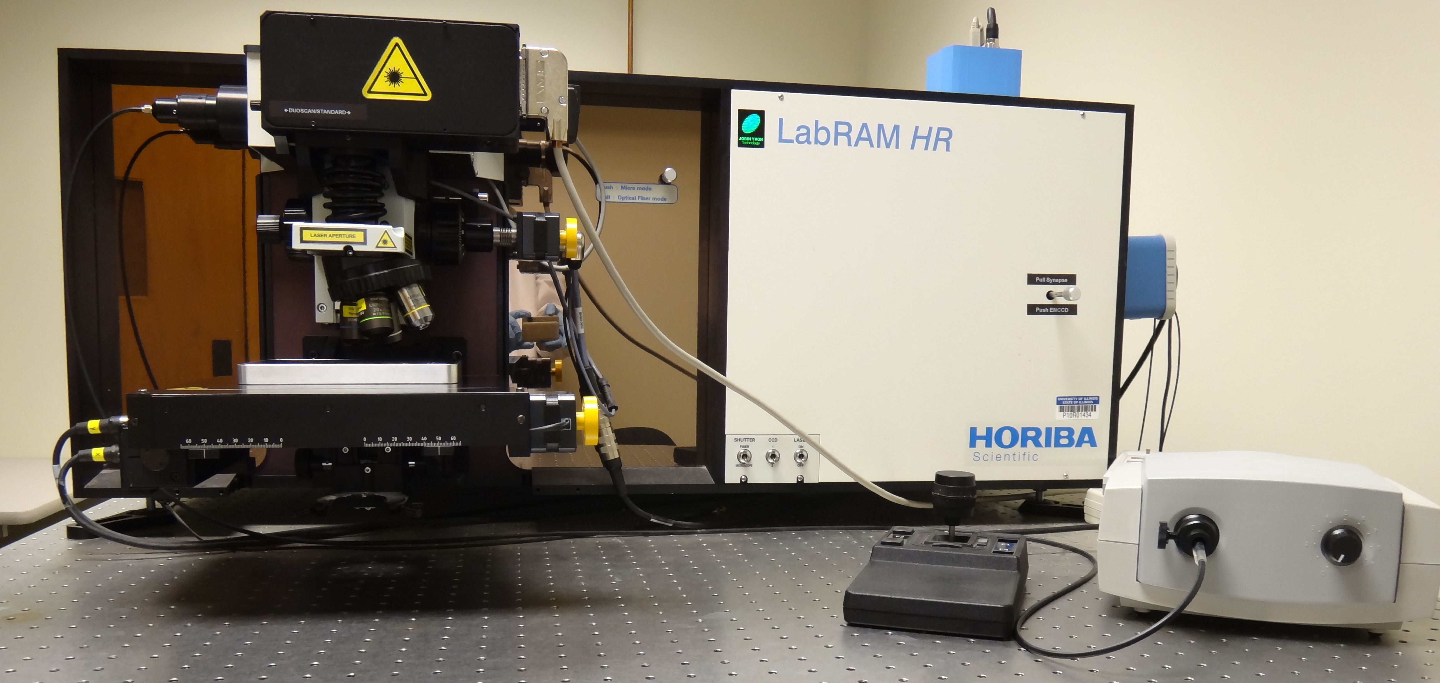
The Raman confocal imaging microscope is a high-resolution research-grade Horiba LabRAM HR 3D-capable Raman spectroscopy imaging system that is optimized for the visible-to-NIR spectral range. It is equipped with four lasers (405, 532, 633, 785, and 830 nm) and has a user-controllable variable aperture confocal pinhole that provides diffraction-limited spatial resolution with maximum signal throughput. A high-precision, high-stability motorized XY mapping stage provides automated control of sample position, with a travelling range of 100 by 80 mm; full Z-axis control is achievable through a Z-axis translation unit with a minimum step size of 0.5 micrometers. A high-speed, computer-controlled visible autofocus system enables stable imaging acquisition over long time periods. The integrated spectrometer has four available gratings and two high-sensitivity cameras: 1) a Horiba Synapse back-illuminated deep-depletion CCD camera and 2) an Andor Newton EMCCD camera. Utilizing the fast Raman imaging technologies SWIFT and DuoScan, this microscope permits the rapid collection of large-area Raman images. The system is suitable for both fluorescence and Raman spectral imaging microscopy, and features are detailed below.
Gratings:
150 g/mm, blazed at 750 nm;
300 g/mm, blazed at 600 nm;
600 g/mm, blazed at 750 nm;
1800 g/mm, blazed at 500 nm.
Objectives:
Visible 10x, NA 0.25, working distance 10.6 mm;
Visible 50x, NA 0.75, working distance 0.38 mm;
Visible 100x, NA 0.95, working distance 0.21 mm;
LWD (long working distance) Visible 20x, NA 0.40, working distance 12 mm;
LWD Visible 50x, NA 0.50, working distance 10.6 mm;
LWD Visible 50x, NA 0.45, working distance 17 mm;
LWD Visible 100x, NA 0.80, working distance 3.4 mm;
LWD Visible 60x (water dipping objective), NA 1.0, working distance 2.0 mm;
LWD NIR 20x (Leica), NA 0.40, working distance 10.7 mm;
LWD NIR 100x (Leica), NA 0.75, working distance 4.7 mm.
Cameras:
Horiba Synapse back-illuminated deep-depletion CCD camera, 1024 x 256 pixels, thermoelectric cooling to -75 Celsius, quantum efficiency >20% over the spectral range of 300 to ~980 nm;
Andor Newton back-illuminated EMCCD camera DU970P, 1600 x 200 pixels, thermoelectric cooling to -100 Celsius, quantum efficiency >20% over the spectral range of ~350 to ~980 nm.
Additional Accessories:
Spectral database management software: Spectral ID searches 1,760 spectral libraries of organics, inorganics, minerals, and other specific types of materials.
Heavy duty computer-controlled x,y motorized stage for use with He Cryostat. The minimum step size is 0.3 micrometers with a traveling range of 102 mm by 102 mm.
The Linkam THMS600 computer-controlled heating and cooling stage operates from -196 °C to 600 °C. This stage has been shared among a few microscopes, so it requires to be booked separately from the microscopes on the calendar.
The Linkam TST350 tensile testing stage operates from -196 °C to 350 °C, with a maximum force of 200 N at a resolution of 0.01 N.
Special Applications:
Duoscan - When the Duoscan application is activated, two scanning mirrors are used to raster the laser beam over the user-determined area, creating one "giant pixel." The size of the giant pixel may be modified using the control software to adjust mirror movement, thus optimizing characterization of large samples.
SWIFT ("Scanning with incredibly fast times") - SWIFT implementation works by scanning the sample stage continuously while reading out the CCD at intervals preset in the software. It enables the acquisition of very large maps of samples where the feature size is much larger than the diffraction-limited resolution of the Raman microscope.
Transmission Raman
Polarized Raman experiments
External fiber port to the spectrometer
SuperHead for 633 nm laser only - Remote spectral measurements
User Resources
- HORIBA Raman Academy - Raman education videos
- Horiba Raman webinars for the Raman confocal microscope
For additional information about this piece of equipment, see the Calendars, Contacts, and Fees pages.
| Primary Contacts | |
|---|---|
| Secondary Contacts | |
| Manufacturer | Horiba |
| Equipment Model | LabRAM HR 3D |
| Location | B606 H |
| Phone Numbers | (217) 333-4387, (217) 244-6928, (217) 265-5071 |
Notes:
* The fee for use is added to our purchase cost for the specific precious metal (gold, gold-palladium, or platinum) expended during coating. Gold and gold-palladium are charged at $0.30/nm, and platinum is charged at $0.45/nm. The added fee is easy to determine for the dual-metal evaporator, for which total thicknesses are reported in nm. For the sputter coaters, at 65 mTorr and 40 mA, gold coats at 3 nm/10 seconds, gold-palladium coats at 1 nm/10 seconds, and platinum coats at 1 nm/10 seconds. So a standard 70-second coat of gold will cost an additional (21 x 0.30) $6.30, a 70-second coat of gold-palladium will cost (7 x 0.30) $2.10, and a 70-second coat of platinum will cost (7 x 0.45) $3.15.