Image Gallery
Gallery
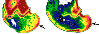
Bone Density Differences Due to Habitual Behavior
Media Details
Created 10/17/2006
These images illustrate a method for inferring differences in habitual knee posture from bone density patterns on the joint surfaces of extant and fossil animals. Both images are oblique slices, obtained from CT data, through the medial femoral condyles (knee joints) of sheep that were trained to walk on treadmills of differing slopes. Color maps were applied using AMIRA software (Mercury Computing Systems) to reflect differences in relative density across the joint surfaces. The subject on the left used a flat treadmill and relatively extended knee postures. The subject on the right used an inclined treadmill and more flexed knee postures. The difference in posture is reflected in their bone properties by the different positions and breadths of the regions of maximum density shown in red. The sheep model was used to validate this methodology and this technique has proved useful for inferring habitual posture and locomotor behavior in extinct subfossil lemurid primates and museum specimens of extant primate taxa. We are in the process of applying this method to infer locomotor patterns used by human ancestors. This work is being conducted in the Evolutionary Biomechanics Laboratory in the Department of Anthropology at the University of Illinois.
Credits
- Dr. John D. Polk , Department of Anthropology