Image Gallery
Gallery
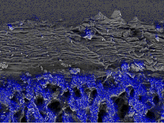
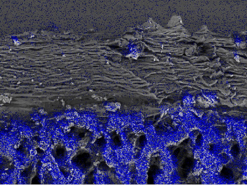
ESEM Image of a Pony Femur
Media Details
Created 4/13/2000
This is a backscattered electron microscopy image of the head of a femur from a young pony. The portions of the image overlaid in blue represent areas in which phosphorus x-rays, indicating the presence of bone, were detected.
Credits
- Scott J. Robinson , ITG, Beckman Institute