Image Gallery
Gallery
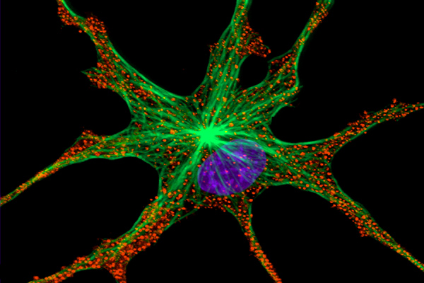
Image of a Xenopus Melanophore

Media Details
Created 6/22/2000
Stained to reveal the microtubule cytoskeleton (green) and the nucleus (blue). The pigment granules in the cell were imaged using back-scattered light. (Confocal Microscope)
Credits
- Steve Rogers