Image Gallery
Gallery
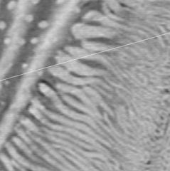
Lamellae in Sea Urchin Tooth
Media Details
Created 1/25/2010
Lamellae between secondary plates in a tooth of the sea urchin Lytechinus variegatus (right portion of the image). Oblique numerical section through 3D synchrotron microCT data set. 1.8 [mu/]m isotropic voxels. SR Stock, X Xiao and F De Carlo obtained the data at station 2-BM of the Advanced Photon Source.
Credits
- Stuart R. Stock , Northwestern University