Image Gallery
Gallery
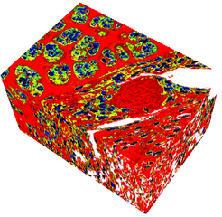
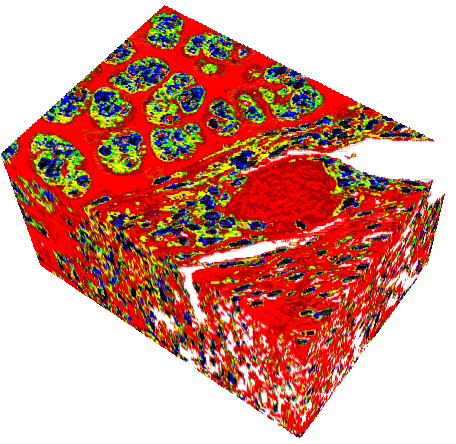
Mouse EHS Sarcoma Tumor Model
Media Details
Created 4/26/2005
The following image is a three-dimensional rendering of an ultrasonic model of a mouse EHS sarcoma tumor. The volume is of size 218 * 156 * 129 micrometer. This volume was obtained from a 43-section optical microscope histologic data set. Among the 43 sections, 7 sections were lost during sectioning at 3 micrometer. Contrast of the sections were equalized and the sections were non-rigidly aligned, while the missing sections were interpolated. A 7-level threshold algorithm was designed to isolate tissue constituents (nuclei, blood vessel, extra-cellular matrix, etc.). Each different color transcribes to assigning different ultrasonic properties (speed of sound, density, acoustic impedance). The volume was used as a computational model for ultrasonic tissue characterization. The digital signal processing algorithms applied to the original images were processed using custom-designed procedures in MATLAB. The data was then imported into Amira and rendered using the Voltex function.
Credits
- Jonathan Mamou , Department of Electrical & Computer Engineering