Image Gallery
Gallery
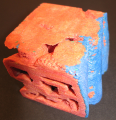
Os-Stained Tissue on a HA Scaffold Revisited
Media Details
Created 2/22/2005
Mouse D1ORL stem cells are cultured on the scaffold for 2 weeks prior to fixation (Os-staining) and micro-CT imaging. Using a raw data histogram, the tissue and the HA can be distinguished based on their x-ray attenuation values. The differences between this image and the Image of the Week for 26 October 2004 lay in the nature of the acquired data, which was made possible using the new SkyScan 1172 MicroCT scanner in the VMIL, and in the processing/analysis of the data, which was accomplished using Amira.
Credits
- Amy Wagoner Johnson , Mechanical Science & Engineering
- Janet Hanlon , ITG, Beckman Institute