Image Gallery
Gallery
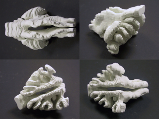
Rodent Nasal Cavity
Media Details
Created 6/22/2004
The model of a rodent's nasal cavity displayed here was derived from a series of histological slices through the head of the rodent. After a subset of the slices were appropriately stained and mounted onto glass slides, they were scanned using the Nikon Super Coolscan 8000 in the ITG. Once completed, the images were stacked and aligned using Photoshop and AnalyzeAVW. The empty space corresponding to the nasal cavity was isolated using the object extraction tools in AnalyzeAVW. The cavity was then converted to a three dimensional surface and printed on the ZCorporation Z406 in the VMIL.
Credits
- Edward Cheng , Molecular & Integrative Physiology
- Esmail Meisami , Molecular & Integrative Physiology