Image of the Week Gallery
Confocal Data Set of Nerve Cell Bodies and Glial Cells

Media Details
Created 4/11/2002
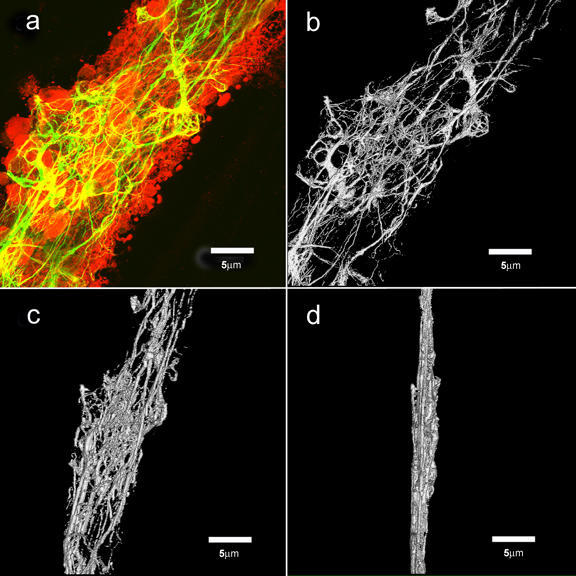
Confocal image stacks of nerve cell bodies and glial cell processes represented in 3-dimensions. Image (a) is a maximum intensity z-projection of the image stack which shows the spatial relationships between differentially labeled cell bodies (Propidium Iodide-red) and glial cell processes (FITC-green). Images (b), (c) and (d) are taken from a volume rendering series of the FITC channel which rotates the structures associated with the glial cells about the y-axis. The z-projection was perfomed using ImageJ; Abramoff's Java implementation (VolumeJ) of the classical object-space rendering algorithm with tri-linear voxel interpolation was used to render the volume. The image data was aquired on the Leica SP-2 spectral confocal and multiphoton microscope housed in the ITG Microscopy Suite.
Credits
- John Chang , Wheeler Laboratory, Neuronal Pattern Analysis Group
- Karl Garsha , ITG, Beckman Institute