Image of the Week Gallery
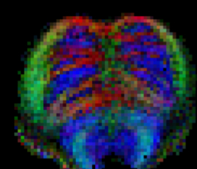
Diffusion Tensor Magnetic Resonance Image of Human Brainstem
Media Details
Created 9/29/2009
High resolution (0.65x0.65 mm2 in-plane resolution) color-coded primary diffusion direction map from diffusion tensor magnetic resonance image of the brainstem on a human subject volunteer, acquired on the 3 T Allegra MRI scanner at the Beckman Institute's Biomedical Imaging Center. Blue indicates superior/inferior, red indicates right/left, and green indicates anterior/posterior directed nerve fiber bundles.
Credits
- Brad Sutton , Bioimaging Science and Technology, Beckman Institute