Image of the Week Gallery
Encapsulation of Parasitic Wasp Eggs
Media Details
Created 11/5/2002
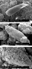
Three ESEM images of parasitic wasp (Cotesia flavipes) eggs, dissected from an unsuitable caterpillar host (Ostrinia nubilalis, the European corn borer). The top image, taken in 'wet' mode, is of an unencapsulated, normal egg, at 4 hours after parasitization. The middle and lower images, taken in 'hi-vac' mode, are of eggs at 8 hours following parasitization. The middle image shows two hemocytes (insect blood cells), the spherical bodies at either end of the egg. The bottom image is of an egg that has been completely encapsulated by hemocytes, which flatten against the egg once they have attached. Other, still spherical, hemocytes - more recent arrivals - are also visible in this image. Encapsulation by the host's immune system will kill the parasitic invaders.
Credits
- Marianne Alleyne , Department of Entomology