Image of the Week Gallery
Hippocampal Neuron Using PC++ Imaging

Media Details
Created 9/9/2008
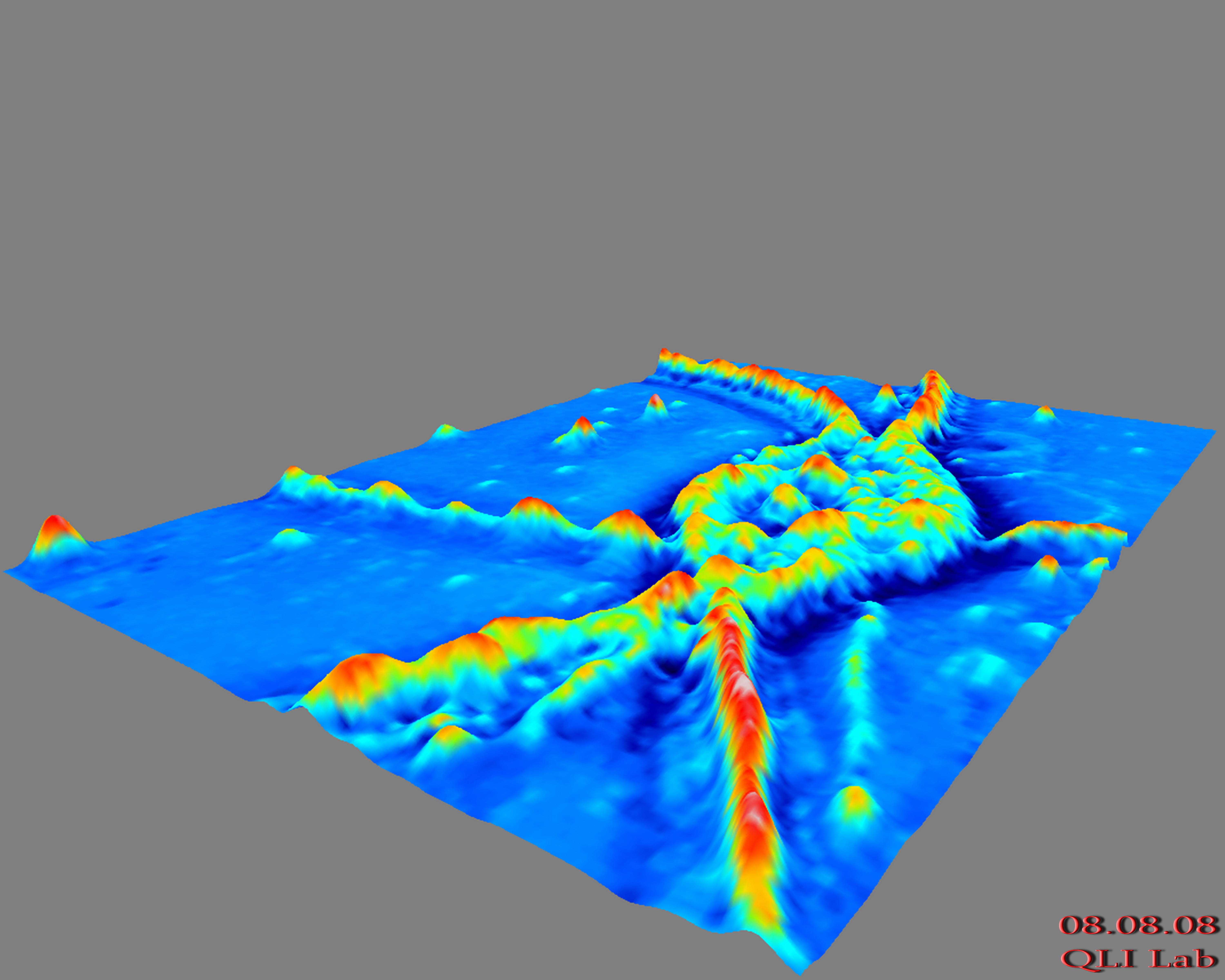
Beckman's Quantitative Light Imaging Laboratory (QLI Lab, http://light.ece.uiuc.edu/), directed by Prof. Gabriel Popescu (Electrical and Computer Engineering and Bioengineering), has developed a novel light microscopy technique aimed at revealing the structure and dynamics of live cells with nanoscale sensitivity and without the need for contrast agents. The new technique, referred to as phase contrast with additional modulation microscopy (phase contrast ++, or PC++) uses interferometry with white light, which brings along significant advantages with respect to lasers. First, the absence of speckle allows for an extremely uniform spatial background profile of less than a nanometer. Second, due to its short coherence length, the white light provides capability for optical sectioning and tomographic imaging of cells. Zhuo Wang from the QLI Lab and Larry Millet from Prof. Martha Gillette's Group (Cell & Developmental Biology, http://www.life.uiuc.edu/clockworks/) apply this ultrasensitive technique to study nanoscale signals in live neurons. This study seeks to reveal new information about intracellular trafficking and cell-to-cell communication. The image shows a live rat hippocampal neuron obtained by PC++. No contrast agents have been used.
Credits
- Zhuo Wang , Bioimaging Science and Technology, Beckman Institute
- Gabriel Popescu , Bioimaging Science and Technology, Beckman Institute