Image of the Week Gallery
Matrix-Assisted Laser Desorption/Ionization Time-of-Flight Mass Spectrometry

Media Details
Created 11/3/2009
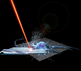
Mammalian neurons from the rat hippocampus and hypothalamus were cultured for direct neuropeptide analysis using mass spectrometry. As graphically depicted here, a crystalline matrix is formed on the cultured neurons to extract and retain the neuropeptides, which are then profiled using Matrix-Assisted Laser Desorption/Ionization Time-of-Flight Mass Spectrometry. This composite image was created using Maya and Photoshop software packages, and produced by collaboration between Larry Millet (Martha Gillette Lab) and Janet Sinn-Hanlon (ITG Visualization Laboratory). The image appears in the article "Direct Cellular Peptidomics of Supraoptic Magnocellular and Hippocampal Neurons in Low-Density Cocultures" by Larry J. Millet, Adriana Bora, Jonathan V. Sweedler and Martha U. Gillette. The paper will be published in the first issue of the online journal "ACS Chemical Neuroscience"; the article is currently available online at http://pubs.acs.org/journal/acncdm. Jonathan Sweedler and Martha Gillette are both faculty in the NeuroTech group at the Beckman Institute.
Credits
- Janet Sinn-Hanlon , ITG, Beckman Institute
- Larry Millet , Neurotech, Beckman Insitute; Department of Cell & Developmental Biology