Image of the Week Gallery
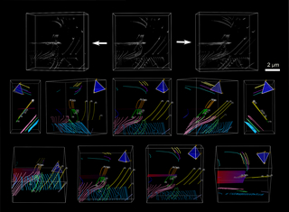
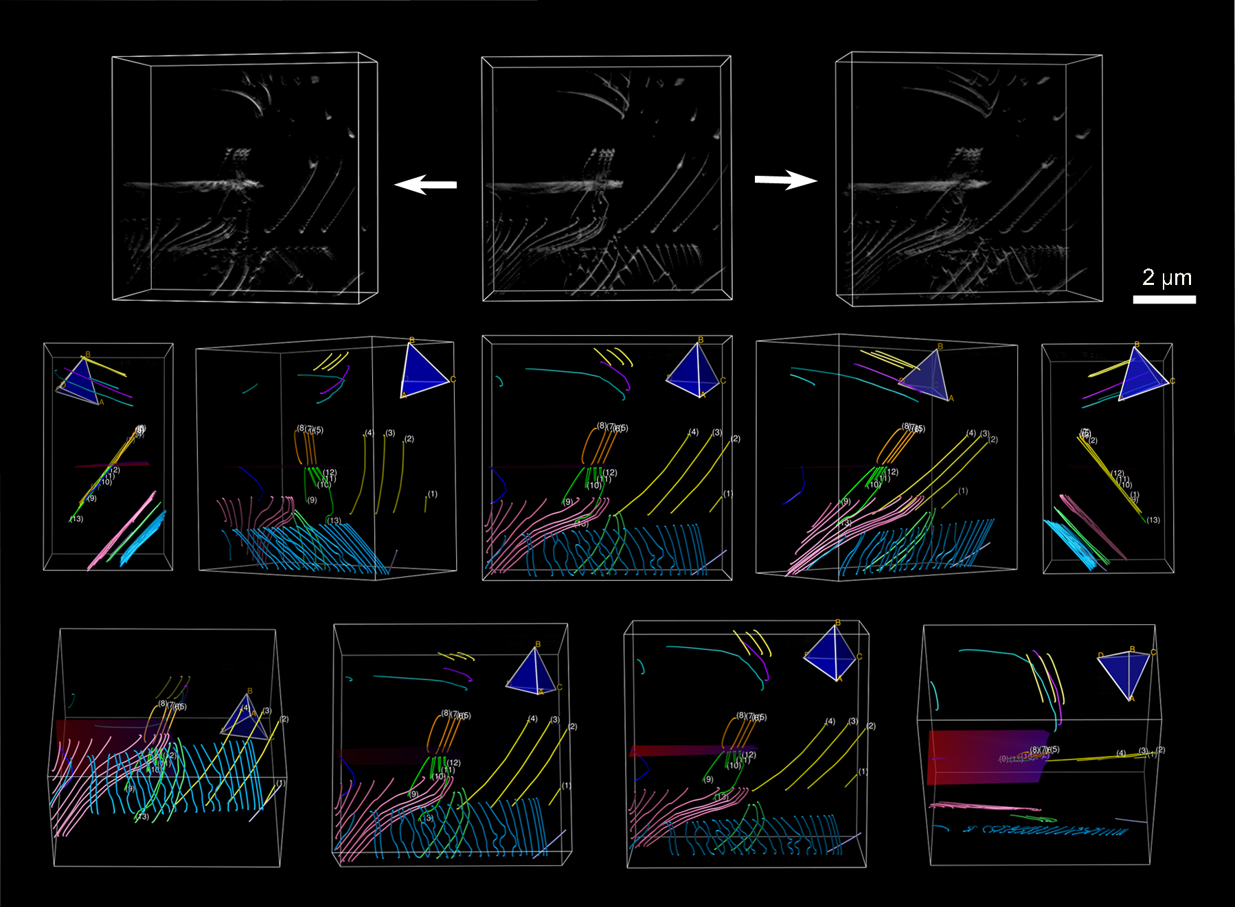
Tomogram Obtained by Transmission Electron Tomography of Dislocation Structures
Media Details
Created 9/28/2010
Top: views from a tomogram reconstructed using bright-field high-voltage electron microscopy (HVEM) images; bottom: views from the traced tomogram, where dislocations were colored according to their slip planes. The crack tip is depicted as a red surface, located middle-left in the volume.
Credits
- Ian Robertson , Materials Science and Engineering
- Kenji Higashida , Kyushu University, Japan
- Stephen House , Materials Science and Engineering
- Grace Liu , Materials Science and Engineering
- Masaki Tanaka , Materials Science and Engineering