Image Gallery
Gallery
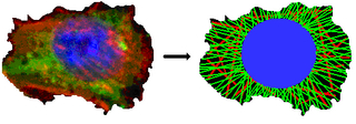
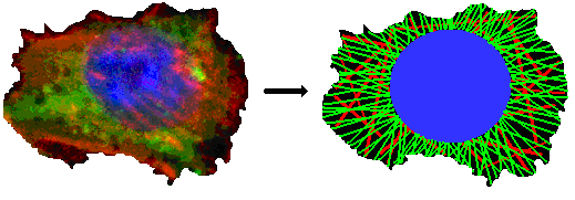
Carcinoma Cell and Simplified Model
Media Details
Created 10/13/2009 5:00:00 AM
Confocal microscope image of stained carcinoma cell (left) and simplified model (right) with nucleus (blue), actin filaments (red), and microtubules (green).
Credits
- Michael Oelze , Bioacoustics Research Laboratory, Beckman Institute
- William D. O'Brien Jr. , Bioacoustics Research Laboratory, Beckman Institute
Imaging Technology Group
405 North Mathews Avenue, Urbana, IL 61801 USA
(217) 300-0566