Image Gallery
Gallery
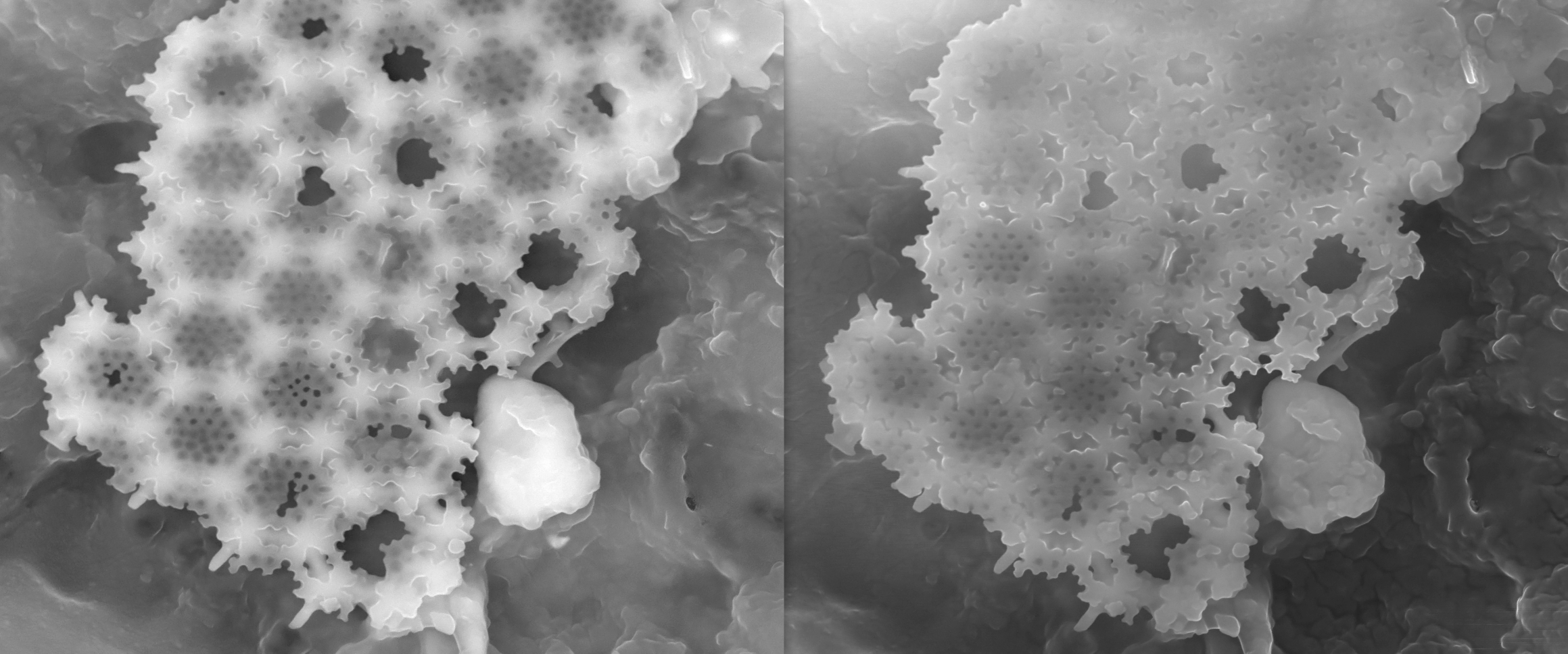
Comparison of Backscatter and Secondary Imaging Using the SEM

Media Details
Created 11/29/2005 6:00:00 AM
These two images, taken at 20 kV with a spot size of 2.6 nm, illustrate the capability of backscattered electron imaging (left panel) to reveal details not readily apparent in secondary electron imaging (right panel). The sample is diatomaceous earth, sometimes used as a fine polishing agent. Original magnification ~10,000x.
Credits
- Aylin Sendemir , Tissue Engineering Group, Beckman Institute
Imaging Technology Group
405 North Mathews Avenue, Urbana, IL 61801 USA
(217) 300-0566