Image Gallery
Gallery
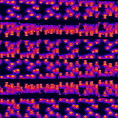
Montage of 3D Animation of Confocal Microscope Point Spread Functions
Media Details
Created 7/18/2002 5:00:00 AM
Fluorescent 190 nanometer diameter polystyrene spheres imaged through laser scanning confocal microscopy (http://www.micromaestro.org animated version). The image illustrates the distortion of axial (z-plane) resolution due to spherical aberration arising from an index mis-match of the mounting media. The spheres were imaged through 20 microns of glycerol-gelatin using a 63x 1.32NA oil immersion objective and 543 nanometer wavelength light. The field of view (each frame) is 740 nanometers.
Credits
- Karl Garsha , ITG, Beckman Institute
Imaging Technology Group
405 North Mathews Avenue, Urbana, IL 61801 USA
(217) 300-0566