Image Gallery
Gallery
Three Dimensional Optical Coherence Tomography of Murine Embryo (E10.5)
Media Details
Created 10/25/2005 5:00:00 AM
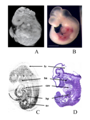
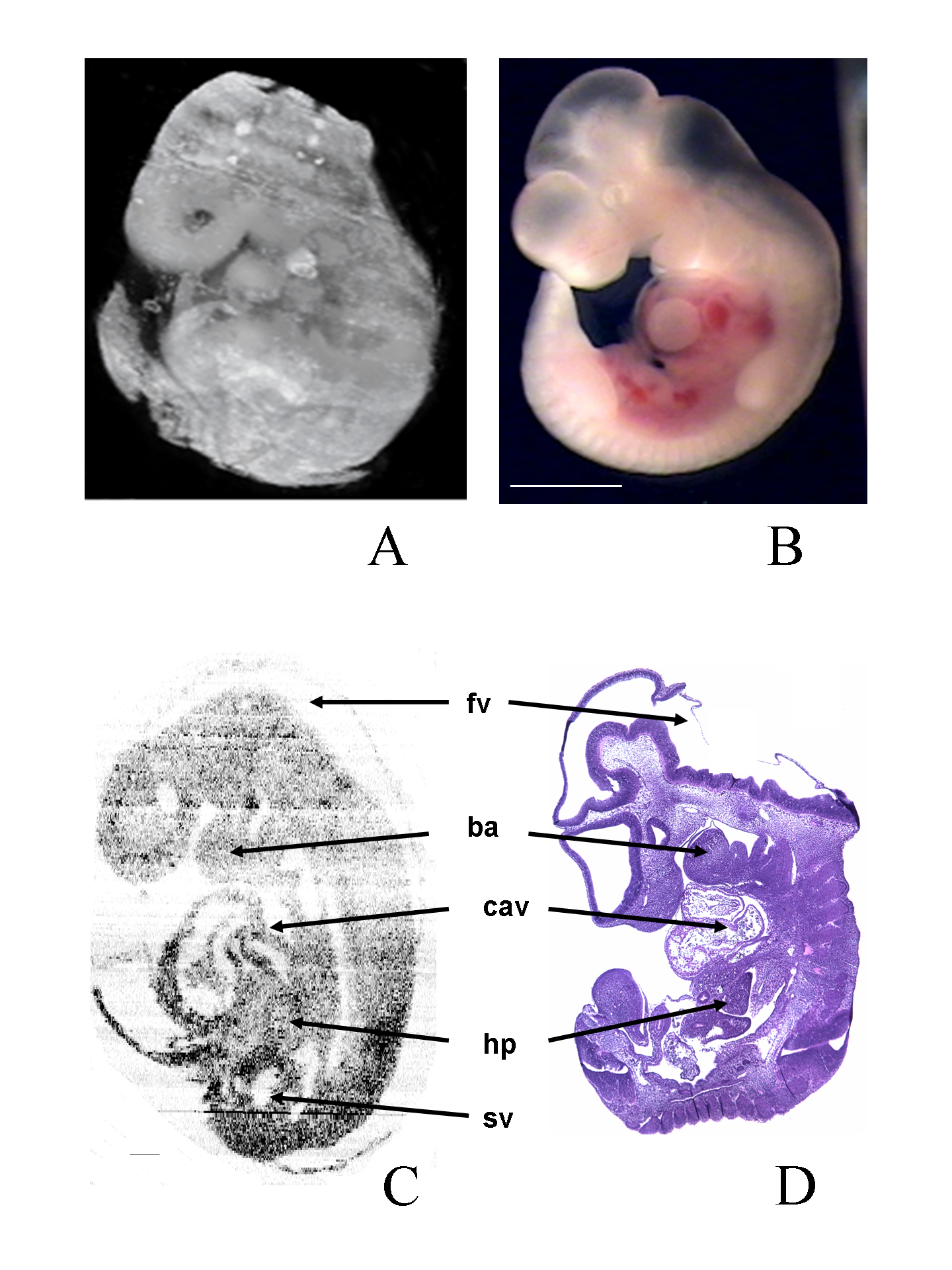
Optical coherence tomography (OCT) is an emerging high-resolution real-time biomedical imaging technology. Figure A is 3D volume rendered by Amira[reg/] software from 256 2D-OCT pictures. Figure B is digital photograph of the E10.5 mouse embryo. Figure C is the computational section at the sagittal plane (3D rendered OCT images from the same data set of fig. A ). Figure D is the correlated histological sections. Ba indicates branchial arch; hp, hepatic primordia; fv, fourth ventricle; sv, subcardinal vein; Scale bar=1mm. Amira is an excellent 3D visualization package available in the VMIL.
Credits
- Janet Sinn-Hanlon , ITG, Beckman Institute
- Dr. Stephen A. Boppart , Biophotonics Imaging Laboratory, Beckman Institute
- Tyler Ralston , Biophotonics Imaging Laboratory, Beckman Institute
- Wei Luo , Biophotonics Imaging Laboratory, Beckman Institute
- Dan Marks , Biophotonics Imaging Laboratory, Beckman Institute
Imaging Technology Group
405 North Mathews Avenue, Urbana, IL 61801 USA
(217) 300-0566