Image Gallery
Gallery
Three Dimensional Reconstructions of Motor Proteins
Media Details
Created 6/29/2000 5:00:00 AM
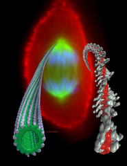
This image represents three-dimensional reconstructions of a microtubule (green) decorated with single headed kinesin (white) and an actin filament (red) decorated with single headed myosin (white). The three-dimensional maps were created using cryo-electron microscopy and helical reconstruction techniques. The maps are shown against a background of a fluorescently stained image of a dividing cell taken on a light microscope. The image was produced using Analyze, SoftImage and Adobe Photoshop in the VMIL.
Credits
- Janet Hanlon , ITG, Beckman Institute
- Abel Lin , Scripps Institute
- Bridget Carragher , ITG, Beckman Institute
- Steve Rogers , ITG, Beckman Institute
- Ron Milligan , Scripps Institute
Imaging Technology Group
405 North Mathews Avenue, Urbana, IL 61801 USA
(217) 300-0566