Details
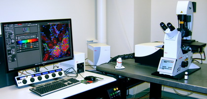
Leica SP8 UV/Visible Laser Confocal Microscope

Confocal microscopy permits one to optically section a fluorescent sample (such as a cell that has been stained with contrasting fluorescent dyes) with superior resolution by using a pinhole to reject light that originates outside of the chosen area. By collecting a series of such images through the depth of a sample, the user may assemble a highly accurate three-dimensional reconstruction of the entire sample.
Features:
- Laser lines: 405, 458, 488, 514, 561 & 633 nm.
- Four detectors: Three PMTs + one high-sensitivity GaAsP HyD detectors.
- Resonant scanning (8kHz) allows 28 fps at 512*512 for fast live cell imaging.
- Scan optics for high transmission from 400 nm to 1300 nm.
- Optical scanfield rotation up to 200 degrees.
- Three mirrors X2Y scanner for parallax-free and tiling imaging.
- Motorized stage, mark & find, tiling confocal imaging.
- LAS X control software.
Objectives:
- 20x/0.75 IMM (oil, water, glycerol) CORR CS2, free Working distance 670 um
- 40x/1.30 HC PL APO Oil CS2, free Working distance 240 um
- 63x/1.40 HC PL APO Oil CS2, free working distance 140 um
Application Modules:
- Fluorescence Recovery After Photobleaching (FRAP).
- Fluorescence Resonance Energy Transfer (FRET)
User Resources
For additional information about this piece of equipment, see the Calendars, Contacts, and Fees pages.
| Primary Contacts | |
|---|---|
| Secondary Contacts | |
| Manufacturer | Leica Microsystems |
| Equipment Model | TCS SP8 |
| Location | B0420B |
| Phone Numbers | (217) 333-4387, (217) 265-5071, (217) 244-6270 (Room B420B) |
Imaging Technology Group
405 North Mathews Avenue, Urbana, IL 61801 USA
(217) 300-0566